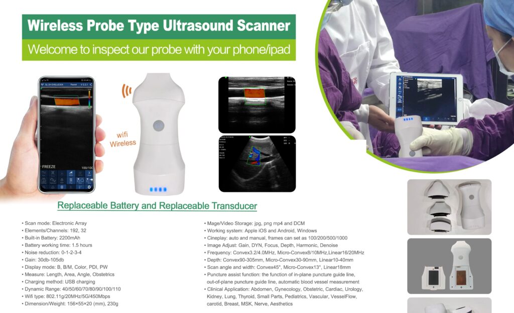
A single wireless probe body accepts seven interchangeable transducer heads — from deep abdominal to fine vascular. Sonographers stop carrying a cart of single-frequency probes and start carrying one case.
About CP-7CPLT
CP‑7CPLT — one handle, seven transducers, every clinical view.
A pocket‑sized WiFi ultrasound probe with a replaceable battery and interchangeable, hot‑swappable transducer heads — from deep abdominal convex to 20 MHz high‑resolution linear and phased‑array cardiac. Streams live B/M/Color/PDI/PW imaging straight to your phone, tablet or Windows PC.
Replaceable battery. Replaceable transducer.
One handle drives seven swap‑in transducer heads, each identified by its frequency range — so a single device covers convex, micro‑convex, linear and phased‑array scanning without buying a new probe.
2.2 / H3.6 MHz
Phased array
Cardiac sector scanning through narrow intercostal windows — echocardiography and adult/paediatric cardiac views.
3.2 / H4.0 MHz
Convex
Deep abdominal and obstetric imaging with a wide 45° field of view.
3.2 / H5.0 MHz
Convex, curved array
General abdomen, kidney, obstetric and gynaecology scanning at extended depth.
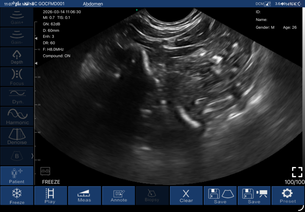
6.6 / H8.0 MHz
Convex
Mid‑range abdominal detail — organ borders and vasculature at 60–90 mm depth.
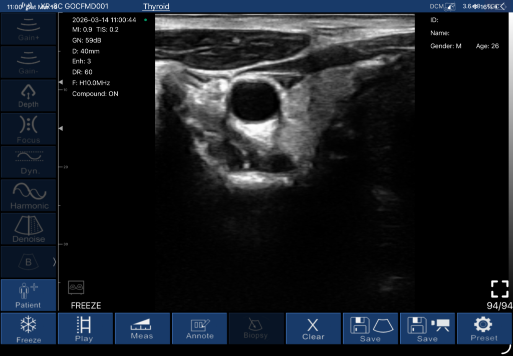
7.5 / H10.0 MHz
Micro‑convex
Thyroid, small parts and pediatric scanning where a compact footprint is needed.
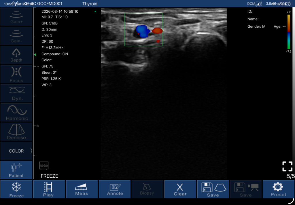
10.0 / H13.2 MHz
Micro‑convex
Higher‑resolution thyroid and superficial vascular imaging with Color Doppler flow.
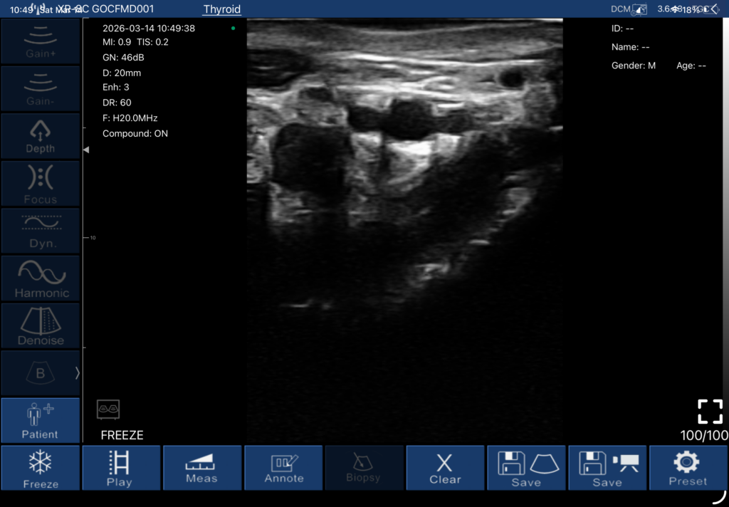
16.0 / H20.0 MHz
Linear array
High‑frequency superficial, nerve, MSK and vascular imaging up to 40 mm depth.
Live scans, straight off the probe
Captured on‑device across the transducer range, streamed to a tablet over WiFi.
Abdomen · Convex 6.6/H8.0 MHz
60 mm depth, tissue harmonic
Thyroid cyst · Micro‑convex 7.5/H10.0 MHz
40 mm depth, B‑mode
Thyroid nodule · Color Doppler 10.0/H13.2 MHz
30 mm depth, red/blue flow mapping
Superficial vessel · Linear 16.0/H20.0 MHz
20 mm depth, high‑resolution B‑mode
See it scanning
Real capture footage from the probe — a high‑frequency linear head resolving a wrist artery, and the phased‑array head sweeping a cardiac view.
Hand / wrist artery imaging
Linear transducer, 18 & 23 MHz
Cardiac scan
Phased‑array transducer
Spec sheet
Bring On-Demand Imaging to Your Clinic
We’d love to hear from you! Drop us a message with your questions, requirements, or feedback, and our team will get back to you soon